
Facilities
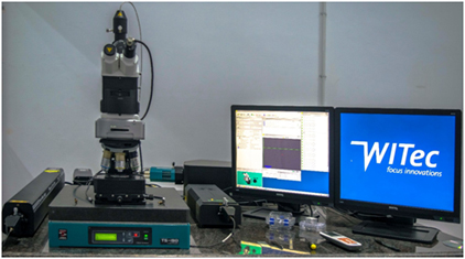
WITEC ALPHA300 RA – CONFOCAL RAMAN MICROSCOPE WITH AFM
Company Name
WITec GmbH, Ulm, Germany
Model Name
Alpha300RA AFM & RAMAN
Spectrograph
UHTS 300, Ultrahigh throughput lens based spectrograph with 300 mm focal length.
CCD
Andor Back illuminated CCD camera with >90% QE in the visible region. The CCD chip is a 1024 x 127 pixels with the pixel size of 24 microns
Description
The instrument is a WITec alpha300RA (WITec GmbH, Ulm, Germany) AFM, SNOM &RAMANcombined system. The laser is coupled into the microscope using a single mode optical fiber coupling and the RAMAN signal is collected into a multimode optical fiber which is connected to the UHTS 300 spectrograph equipped with a back illuminated CCD with better than 90% QE in the visible region.
Laser
532 nm DPSS laser with a maximum power after single mode fiber of around 70 mW.
Gratings
UHTS 300 is equipped with a motorized turret that can mount 3 gratings. The gratings available with the instrument are 600 g/mm BLZ 550 nm & 1800 g/mm BLZ 550 nm.
Optical Microscope
Modified Zeiss Axioscope.
Objectives
100X Zeiss 0.9 NA, 50X Zeiss 0.75 NA, 20X Zeiss 0.4 NA, 10X Zeiss 0.25 NA
Working
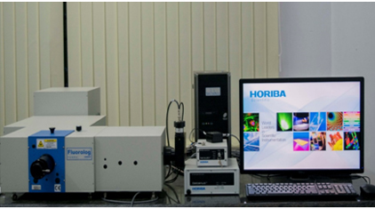
HORIBA FLUOROLOG FLUOROSCENCE SPECTROMETER COUPLED WITH INTEGRATING SPHERE AND LIFETIME DETECTOR
Company Name
Horiba, USA
Model Name
Fluorolog 3 TCSPC
Excitation sources Steady-state
Broadband 450-W xenon arc lamp from UV to near-IR.
Excitation sources TCSPC
295,330,570nm-NanoLed
355,395nm-SpectralLed
Spectrometers
FL3-21, Single-grating emission and double-grating excitation monochromator.
Grating
1200-grooves/mm
Resolution
0.2 nm
Accuracy
±0.5 nm
Speed
150nm/s
Range
0-1300nm
Detector
RT298P (PMT)
Lifetimes measurable
< 100 ps to 100 microsecond lifetimes
Working
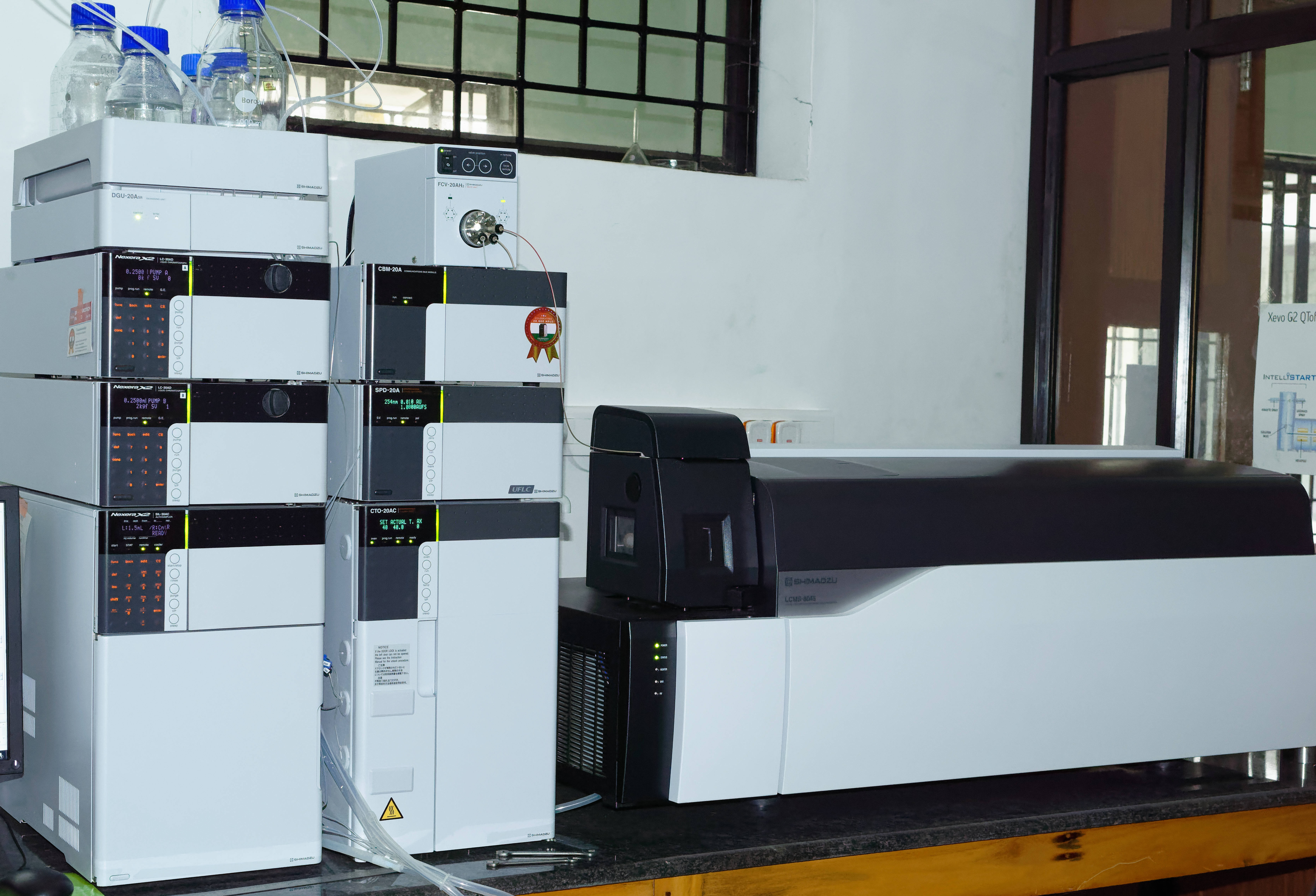
LIQUID CHROMATOGRAPHY- MASS SPECTROMETER (LC-MS/MS)
Company Name
Shimadzu
Model Name
8045
Interface
ESI and APCI
Analysis Modes
Q1 Scan/SIM
Q3 Scan/SIM
Precursor ion Scan
Product ion scan
Neutral loss scan
Q3 Scan/SIM
Precursor ion Scan
Product ion scan
Neutral loss scan
Detector
Secondary Electron Multiplier with off-axis conversion dynode
Working
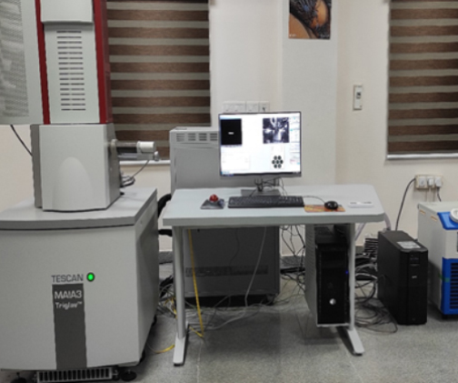
FIELD EMISSION SCANNING ELECTRON MICROSCOPE (FE-SEM) WITH EDS
Company Name
TESCAN BRONO s.r.o.Czech
Model Name
MAIA3 XMH
About
MAIA3 – an exceedingly powerful tool for comprehensive nano materials characterization, gentle observation of beam-sensitive samples and comfortable imaging of non-conductive samples including uncoated biological specimens and semiconductors. The finest sub-nanometer details of even low-contrast samples are captured using the Triglav™ SEM column along with the ultra-high resolution Trilens™ optics and an impeccable detection system with triple SE and triple BSE detectors. Userfriendliness is enhanced by a large number of sophisticated SW modules and automatic procedures.
MAIA 3 model was designed to perform an imaging with low-energy electrons and an ultra-high resolution at the same time.
The low energy electrons give better material contrast, prevent specimens from possible damage and non- conductive specimens from charging.
It is having XMH microscope which is working only in high vacuum conditions.
In high vacuum mode it is possible to investigate both conductive and non-conductive samples ( non-conductive samples require previous metal (sputtered) or carbon coating.)
Essential Specifications & Features
Electron Gun: High brightness Schottky emitter
Resolution: 0.7 nm at 15 kV
1.0 nm at 1 kV (with BDM Mode)
Magnification: 4x to 1,000,000x
Maximum Field of View: 4.3 mm at WD analytical 5 mm
7.5 mm at WD 30 mm
Accelerating Voltage: 200 V to 30 kV/ / down to 50 eV with
BDT
Probe Current: 2 pA to 400 Na
Specimen Stage: Compucentric, fully motorized Movements
Electron Optics Working Modes:
Resolution: Ultra-high resolution mode
Depth: Sets the column up in a mode that enhances depth of focus
Field: Optimizes the column to provide a large non-distorted field of view, field-free mode suitable for magnetic samples
Detector
SE Detector
* In-Beam SE Detector
* BSE Detector
* In-Beam BSE Detector
* In-Beam SE Detector
* BSE Detector
* In-Beam BSE Detector
Working